A leap forward for in vivo histology
Virtual chromoendoscopy with i-scan
The i‑scan technology provides extra visual information for a more accurate in vivo diagnosis through improved vessel and mucosal pattern characterization.

Different i‑scan modes
We introduced i‑scan to offer digital image processing on all our video processors. The INSPIRA and OPTIVISTA video processors are additionally equipped with i‑scan Optical Enhancement (OE) which adds bandwidth-limiting light for further improved visualization.
i‑scan
The digital enhancements consist of three different image algorithms:
Surface Enhancement (SE)
Tone Enhancement (TE)
Contrast Enhancement (CE)
All i‑scan modes can be applied individually or in combination with one another.
Switching the levels or modes of enhancement can be performed on a real‑time basis, without any time lag, by pushing an endoscope button, enabling efficient endoscopic observation.

Surface enhancement
The i‑scan SE mode enhances the mucosal structure in a natural color tone to support detection, especially of flat lesions by highlighting abnormalities.

Tone Enhancement
The i‑scan TE mode enhances the changes of vascular and mucosal structure with a color tone change, supporting pattern characterization.

Contrast Enhancement
The i‑scan CE mode enhances depressed areas and differences in structure by adding blue color to the edges.
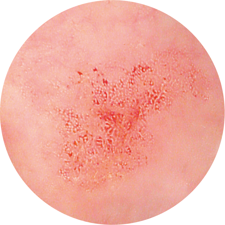
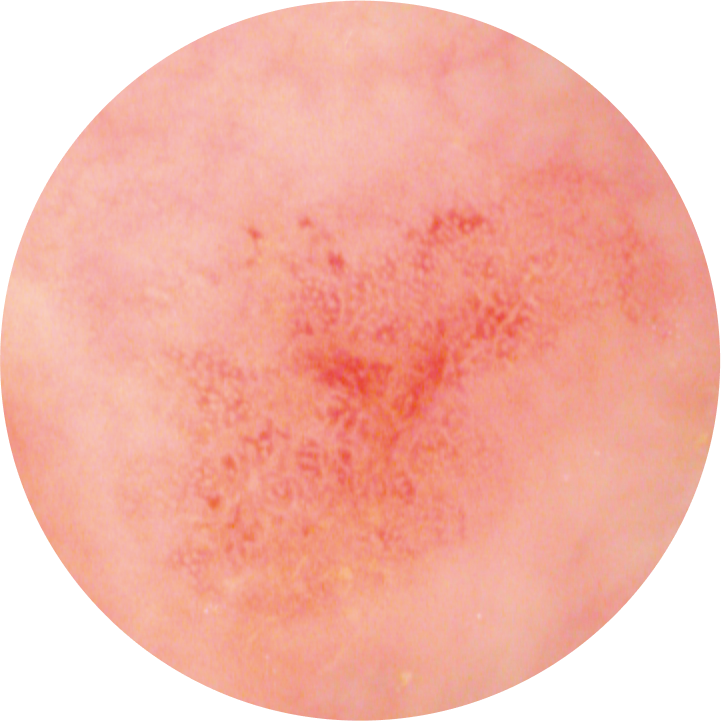
Detection support
i‑scan SE is proven to enhance lesion detection by 11%. SE enhances light‑dark contrast, which in turn enhances the structure through recognition of the edges. It retains the natural white light color tones and thus can be kept activated during the entire procedure.
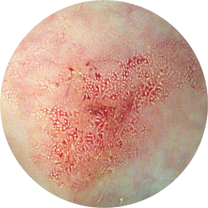
Pattern characterization
i‑scan TE enhances both pit pattern and vascular structures in a color tone that contributes to characterization and demarcation. TE dissects and analyzes the individual RGB components of a normal image and enhances minute mucosal structures and subtle changes in color.
Optical enhancement
i‑scan OE combines bandwidth-limiting light with digital image processing technology.
This allows for higher contrast display of the surface structures of blood vessels, glandular ducts and mucosal membranes compared to white light.
i‑scan OE is designed to support physicians to improve characterization, detection and diagnosis of gastrointestinal lesions more precisely.
i‑scan OE supports the screening and detailed examination of lesions using two modes:

Mode 1
Provides sufficient light and highlights blood vessels.
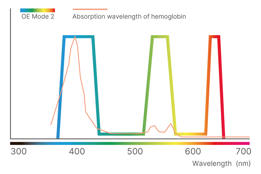
Mode 2
Enhances the blood vessels and mucosa in natural white light, potentially advancing screening procedures.
Twin Mode
PENTAX Medical's unique Twin Mode functiondisplays a normal white light image and an i‑scan image simultaneously, side by side. Switching between the white light and i‑scan modes can be done in real time.
The Twin Mode function aids in both routine, everyday procedures and in educational applications.