PENTAX i‑SCAN™
Image Enhancement
Alongside the advancement of high-resolution endoscopes, endoscopic adjunctive techniques and technologies have been developed. These recent technological innovations are focused on assisting the endoscopist better visualize images through various enhancements of high-definition white light endoscopy (HDWLE).
i-SCAN Defined
i-SCAN is a digital, post-processing image enhancement technology that provides the user an enhanced view of the texture of the mucosal surface and blood vessels. i-SCAN has three default settings: i-SCAN 1, i-SCAN 2, and i-SCAN 3, all of which can be simply accessed with the touch of a button. Switching between HDWLE and the three i-SCAN settings is quick and can be initialized on a real-time basis.
i-SCAN: How does it work?
Powered by the new PENTAX Medical EPK-i5010 Video Processor, i-SCAN functions by performing per-pixel modifications of the white light image.
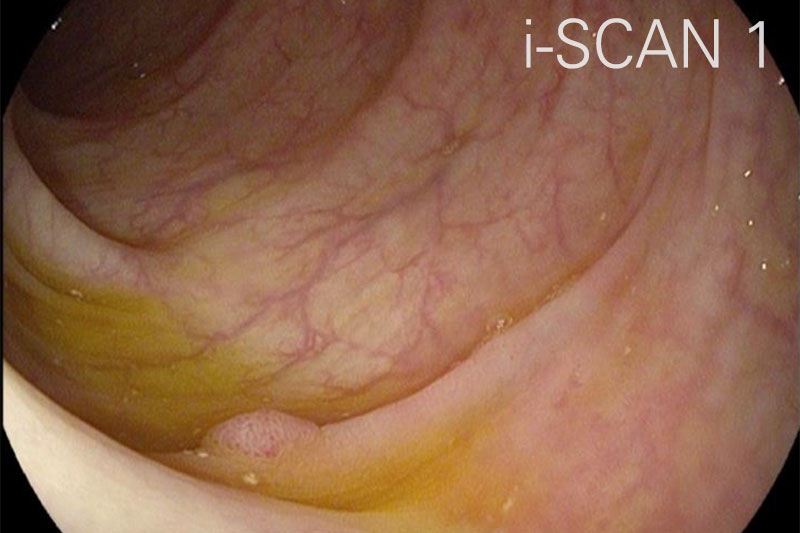
- i-SCAN 1, comprised of surface enhancement and contrast enhancement, provides the user with a view that sharpens surface vessels and enhances surface texture of the mucosa.
- i-SCAN 2, comprised of surface enhancement, contrast enhancement, and tone enhancement mode c, provides the user with an image that increases the contrast between the mucosa and blood vessel. This increased contrast leads to improve visibility of blood vessels while also providing the same enhancements to the mucosal surface texture achieved in i-SCAN 1.
- i-SCAN 3 (surface enhancement, contrast enhancement and tone enhancement mode g) provides the user with increased visibility of blood vessels including dimly illuminated far-field regions, while also providing the same enhancement to the mucosal surface texture achieved in i-SCAN 1.
