Virtual Chromoendoscopy With i-scan
The i‑scan technology provides extra visual information for a more accurate in vivo diagnosis through improved vessel and mucosal pattern characterization.

Different i‑scan Modes
i‑scan consists of three different image algorithms that are arranged in series, therefore, it is possible to apply two or more of these modes at one time. Switching the levels or modes of enhancement can be performed on a real‑time basis, without any time lag, by pushing a scope button, enabling efficient endoscopic observation.
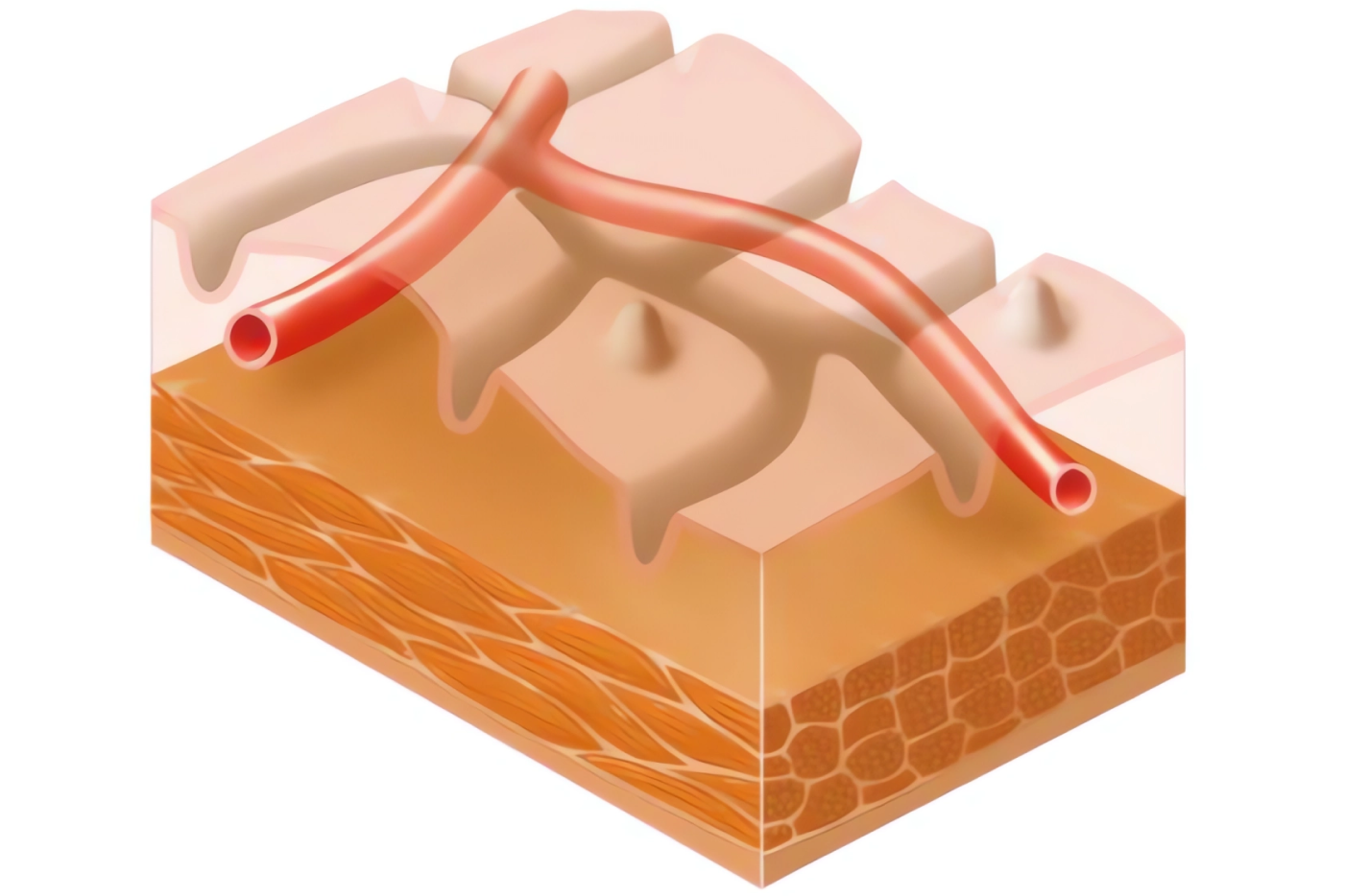
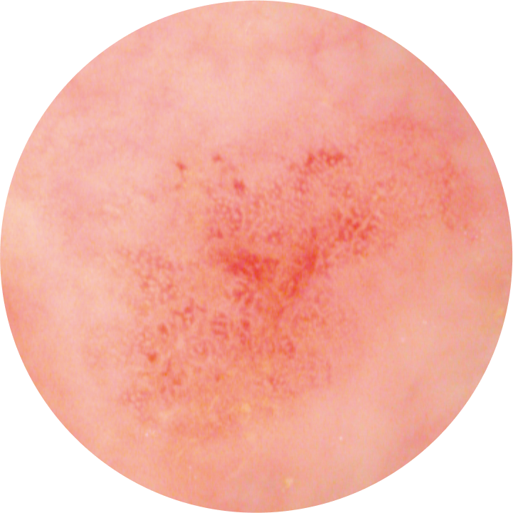
Surface Enhancement
The i‑scan SE mode enhances the mucosal structure in a natural color tone to support detection, especially of flat lesions by highlighting abnormalities.
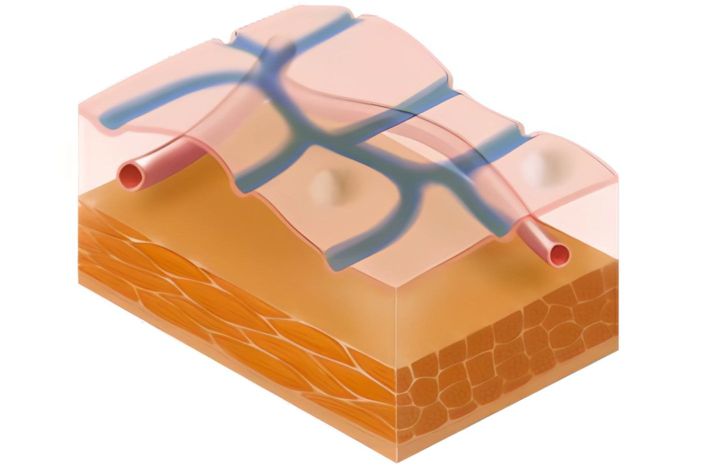
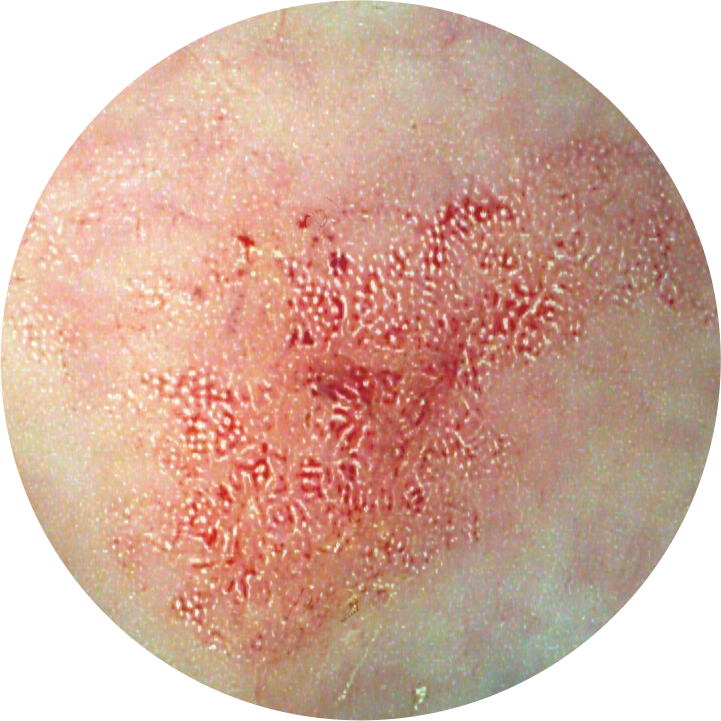
Tone Enhancement
The i‑scan TE mode enhances the changes of vascular and mucosal structure with a color tone change, supporting pattern characterization.
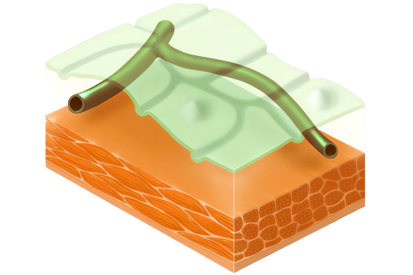
Contrast Enhancement
The i‑scan CE mode enhances depressed areas and differences in structure by adding blue color to the edges.
Detection
i‑scan SE enhances light‑dark contrast which in turn enhances the structure through recognition of the edges. It retains the natural color tones thus can be kept activated during the entire procedure.
Pattern Characterization
i‑scan TE enhances both pit pattern and vascular structures in a color tone that contributes to characterization and demarcation. TE dissects and analyzes the individual RGB components of a normal image and enhances minute mucosal structures and subtle changes in color.
Optical Enhancement
PENTAX Medical has developed an Optical Enhancement (OE) function to further improve the visibility of blood vessels, gland ducts, and surface structures.
The OE function combines bandwidth-limiting light with digital image processing technology. This allows for higher contrast display of the surface structures of blood vessels, glandular ducts, and mucosal membranes compared to white light. This combination has the potential to help physicians improve characterization, detection, and diagnosis of gastrointestinal lesions more precisely.
The OE filter supports the screening and detailed examination of lesions using two modes. Mode 1 provides sufficient light and highlights blood vessels, while Mode 2 enhances the blood vessels and mucosa in natural color, potentially aiding in screening.

Twin Mode
PENTAX Medical’s unique Twin Mode functiondisplays a normal white light image and i‑scan image simultaneously side by side. Switching between the white light image and three i‑scan settings can be done in real time.