.jpg)
i‑scan imaging
PENTAX Medical i-scan image processing provides digital image enhanced endoscopy (IEE). i-scan provides real-time virtual chromoendoscopy1 for a detailed view of mucosal and vascular patterns with three different enhancement options that highlight specific anatomical features. This range of enhanced visualization modes can support early detection, demarcation and characterization of disease lesions. Together with PENTAX Medical’s advanced HD endoscopes, i-scan has been proven to increase adenoma detection rates to help physicians meet quality measurement standards.1,2
Control what you see with enhanced options
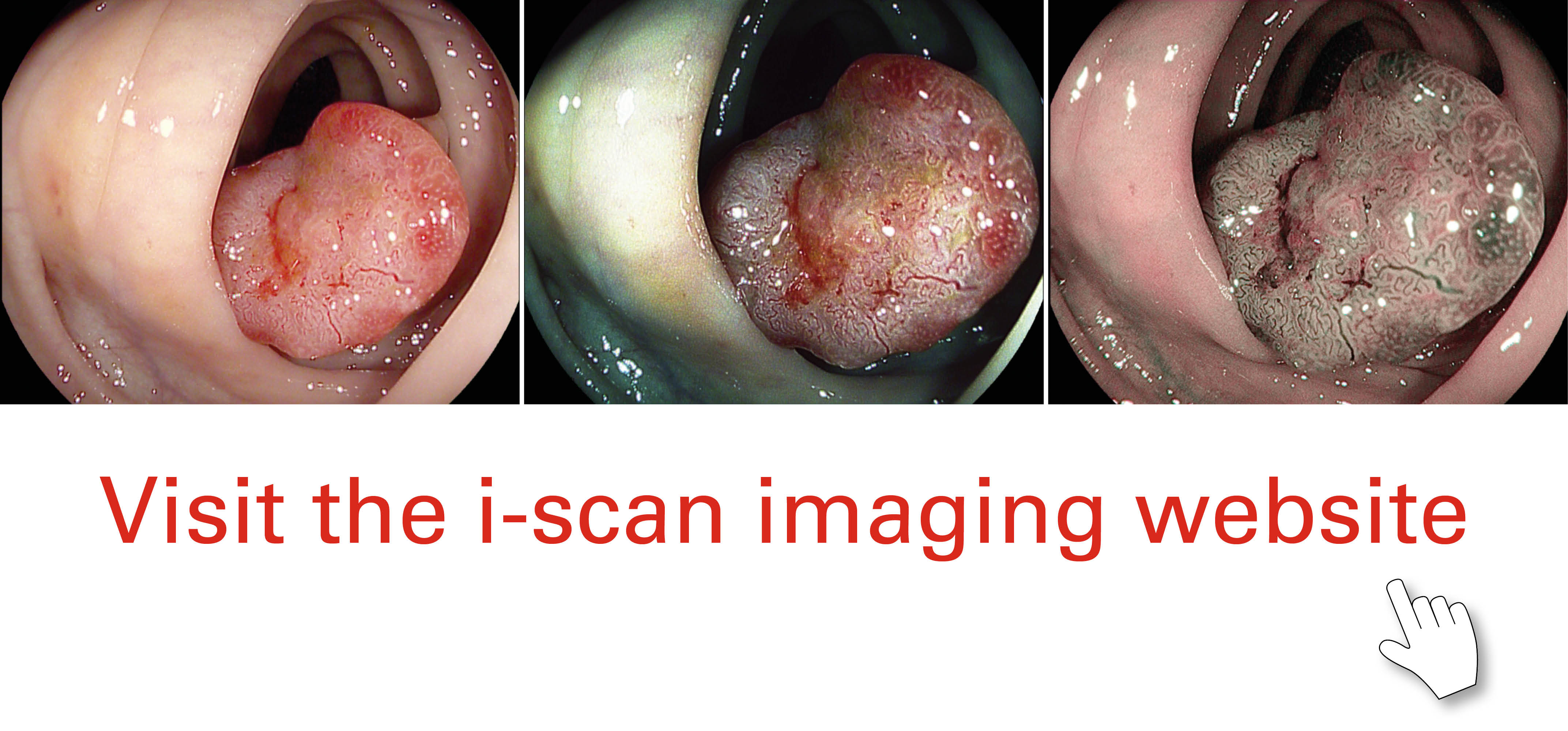
i-scan image processing includes three algorithms: Surface Enhancement (SE), Contrast Enhancement (CE), and Tone Enhancement (TE). Physicians can adjust visualization as needed and switch seamlessly between the modes to view different aspects of suspected disease. SE can help visualize the edges of anatomical structures; CE can help visualize depressed areas through colored presentation of low-density areas; and TE can help tailor enhancement by modifying the colorization of each pixel. PENTAX Medical’s unique Twin Mode function (available on EPK-i7000 video processors) displays a normal white light image and i-scan image simultaneously side by side. Switching between the white light image and three i-scan settings can be done in real time.
Enhanced visualization, improved detection
i-scan real-time image processing provides visual enhancement that can improve disease detection. SE can better delineate mucosal structure and tissue folds, making structures appear elevated and blood vessels accentuated. CE can sharpen the appearance of surface vessels and enhance the visualized details of mucosa surface texture. Both SE and CE retain natural color tones and can be used during the entire procedure. TE accentuates mucosal patterns and vascular structures to aid in lesion characterization. Together with PENTAX Medical HD endoscopes, i-scan image enhancement has been proven to increase adenoma detection rates.1,2
References
- Neumann H, Vieth M, Fry LC., et al. Learning curve of virtual chromoendoscopy for the prediction of hyperplastic and adenomatous colorectal lesions: a prospective 2 center study. Gastrointestinal Endoscopy. 2013;78(1):115-120.
- Hong SH, Choe WH, Lee JH, et al. Prospective, randomized, back-to-back trial evaluating the usefulness of i-SCAN in screening colonoscopy. Gastrointestinal Endoscopy. 2012;75(5): 1011-1021.
Image courtesy of Prof. Helmut Neumann, Professor of Molecular Endoscopy, Department of Medicine 1, University of Erlangen-Nuremberg, Erlangen, Germany (colorectal polyps) and Dr. Matthew Banks, Dr. Rehan Haidry, Dr. Laurence Lovat, Gastroenterologists, University College Hospital, London, UK (Barrett's oesophagus)
Explore more: The i-scan imaging project
The i-scan imaging project is a collaborative initiative led by PENTAX Medical, working with with Key Opinion Leaders from throughout the endoscopic community. It aims to inform endoscopists of this exciting new technology and promote its use in routine clinical practice, helping to improve the quality of diagnostic endoscopy.